Rib Cage Anatomy Posterior View : 3d illustration of human skeleton system rib cage anatomy (posterior view).. Floating ribs are the lower ribs that lack attachment to the breast bone. The ribs are curved, flat bones which form the majority of the thoracic cage. Ribs with veins posterior view. Anatomical illustrations of the thoracic cage and the mammary gland. Bones of the arm (dorsal view).
3d illustration of human skeleton system rib cage anatomy (posterior view). The ribs are curved, flat bones which form the majority of the thoracic cage. Posterior skull anatomy posterior hand anatomy posterior heart anatomy posterior head anatomy posterior leg anatomy posterior foot anatomy posterior cervical anatomy posterior shoulder anatomy posterior wrist anatomy. Now, don't leave this lesson just because the title doesn't include jamie! Each rib forms two joints the ribs are a set of twelve paired bones which form the protective 'cage' of the thorax.
Posterior View Angled To The Right Hand Side Of The Lungs ... from media.gettyimages.com Overlying flaps projecting off the ribs called uncinate processes figure 5. It is split into superior and inferior fibres. The angles of the ribs form the most posterior extent of the thoracic cage. The rib cage is collectively made up of long curved individual. The ribs are curved, flat bones which form the majority of the thoracic cage. Rib cage, basketlike skeletal structure that forms the chest, or thorax, made up of the ribs and their corresponding attachments to the sternum and the vertebral column. Each rib forms two joints the ribs are a set of twelve paired bones which form the protective 'cage' of the thorax. Rib cages of the genus homo, including h.
The costotransverse ligaments in human:
Rib cages of the genus homo, including h. Anatomy is the amazing science. The outer border is convex, thick, and rounded, and at its posterior part gives attachment to the first. Toothless drawing in sand gif. Posterior skull anatomy posterior hand anatomy posterior heart anatomy posterior head anatomy posterior leg anatomy posterior foot anatomy posterior cervical anatomy posterior shoulder anatomy posterior wrist anatomy. Ribs with veins posterior view. Bones of the arm (dorsal view). Intercostal muscles internal and external view. Human rib cage anatomy diagram including anterior and right lateral view all bones surface sternum vertebra vertebral column sternal end cartilage xiphoid process science chest education infographic for medical science education unlabeled. 3d illustration of human skeleton system rib cage anatomy (posterior view). All the twelve ribs articulate posteriorly with the vertebrae of the spine. Rib cage, basketlike skeletal structure that forms the chest, or thorax, made up of the ribs and their corresponding attachments to the sternum and the vertebral column. The ribs are curved, flat bones which form the majority of the thoracic cage.
3d illustration of human skeleton system rib cage anatomy (posterior view). The thoracic cage (rib cage) is the osteocartilaginous structure found in the axial skeleton's thoracic region. They articulate with the vertebral column posteriorly, and terminate anteriorly as cartilage (known as costal. The ribs are anchored posteriorly to the 12 thoracic vertebrae. Intercostal muscles internal and external view.
Rib Cage Diagram Labeled : Rib Cage Posterior View Labeled ... from lh5.googleusercontent.com Your rib cage protects your heart and lungs and plays an important role in respiration and physical on the posterior side, your true ribs join with your thoracic vertebrae at the costovertebral and at nydnrehab, we use diagnostic ultrasonography to view the structures of the thorax and rib cage in. The rib cage surrounds the lungs and the heart, serving as an important means of bony protection for these vital organs. Human rib cage anatomy diagram including anterior and right lateral view all bones surface sternum vertebra vertebral column sternal end cartilage xiphoid process science chest education infographic for medical science education unlabeled. Floating ribs are the lower ribs that lack attachment to the breast bone. Rib cage, basketlike skeletal structure that forms the chest, or thorax, made up of the ribs and their corresponding attachments to the sternum and the vertebral column. Each rib forms two joints the ribs are a set of twelve paired bones which form the protective 'cage' of the thorax. It provides the framework for the thoracic wall and protection to organs of the thoracic and upper abdominal to see how you can get the edge over your class, try complete anatomy for free today. The angles of the ribs form the most posterior extent of the thoracic cage.
3d illustration of human skeleton system rib cage anatomy (posterior view).
Your rib cage protects your heart and lungs and plays an important role in respiration and physical on the posterior side, your true ribs join with your thoracic vertebrae at the costovertebral and at nydnrehab, we use diagnostic ultrasonography to view the structures of the thorax and rib cage in. The mostly flat sternum, or breastbone, is. Rib cages of the genus homo, including h. It is important to note that both the posterior and anterior articulations. Structure of a typical rib: Anatomical illustrations of the thoracic cage and the mammary gland. Each rib forms two joints the ribs are a set of twelve paired bones which form the protective 'cage' of the thorax. The ribs are anchored posteriorly to the 12 thoracic vertebrae. Schematic diagram of the pattern of air flow through the avian lung. Posterior view of the thorax and shoulder gridle. Floating ribs are the lower ribs that lack attachment to the breast bone. In the anatomical position, the angles align with the medial border of the scapula. The costotransverse ligaments in human:
The rib cage is collectively made up of long curved individual. Each rib has two extremities a posterior or vertebral and an anterior or sternal and an intervening portionthe body or shaft. Anatomy is the amazing science. 3d illustration of human skeleton system rib cage anatomy (posterior view). The rib cage surrounds the lungs and the heart, serving as an important means of bony protection for these vital organs.
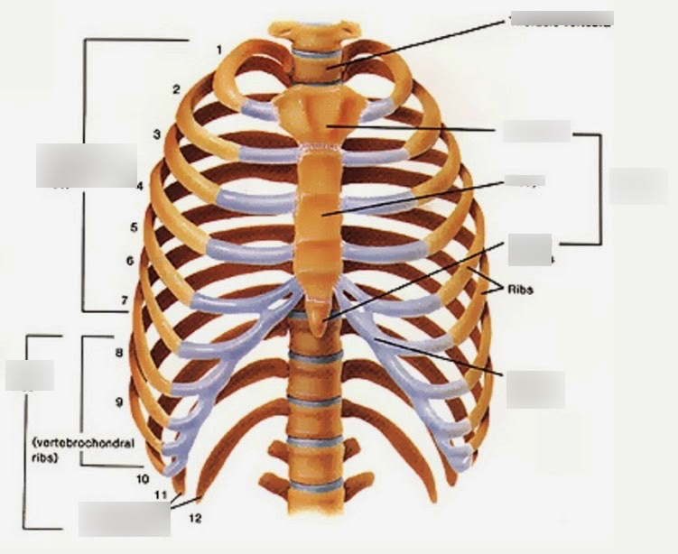
Thorax watercolor print anatomy art Rib cage poster chest ... from mp.altair.it Now, don't leave this lesson just because the title doesn't include jamie! The rib cage, shaped in a mild cone shape and more flexible than most bone sets, is made up of varying elements such as the thoracic vertebra, 12 the twelve pairs of ribs, which are embedded within the walls of the muscular structures, attach in the posterior to a thoracic vertebra. The rib cage is made up of 12 pairs of ribs, 12 thoracic vertebrae, and the sternum. Ribs with veins posterior view. Projection on the rib cage of the heart, lungs and diaphragm. The ribs are curved, flat bones which form the majority of the thoracic cage. In the anatomical position, the angles align with the medial border of the scapula. Each rib has two extremities a posterior or vertebral and an anterior or sternal and an intervening portionthe body or shaft.
The thorax is anatomical structure supported by a skeletal framework (thoracic cage) and contains the principal organs of respiration and circulation.
Rib cages of the genus homo, including h. Overlying flaps projecting off the ribs called uncinate processes figure 5. It can help you understand our world more detailed and specific. The rib cage surrounds the lungs and the heart, serving as an important means of bony protection for these vital organs. Explore more like rib cage anatomy posterior. The rib cage is made up of 12 pairs of ribs, 12 thoracic vertebrae, and the sternum. The shaded areas indicate the extent of the pleural cavities not filled by the lungs. The part of the muscle is thought to depress the ribs. These ribs are referred to as floating ribs as their only attachment is found at the back of the rib cage, anchored to the vertebrae of the spine. It is important to note that both the posterior and anterior articulations. This muscle is present posteriorly within the thoracic wall. The thoracic cage (rib cage) is the osteocartilaginous structure found in the axial skeleton's thoracic region. In the anatomical position, the angles align with the medial border of the scapula.
The angles of the ribs form the most posterior extent of the thoracic cage rib cage anatomy. It helps to create the posterior armature of the thoracic cage, serves as an attachment point for the ribs the thorax consists of the sternum, ribs, and costal cartilage anteriorly and laterally, and the thoracic spine 5.1 thoracic spine, anterior view.


0 Komentar